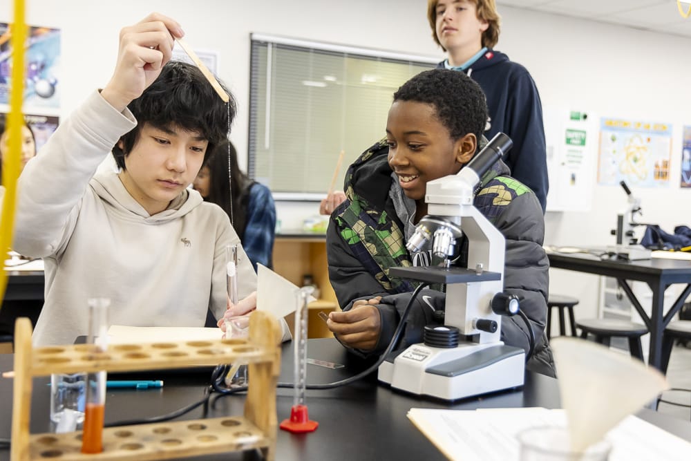
Did you know Dutch eyeglass makers invented the first microscopes in the late 1500s? However, it wasn’t until the late 1700s that two researchers made the microscope an official scientific instrument. In the Middle School Winter Term Microscopy course, taught by Science Teacher William Bander, students got a first-hand view by exploring the parts and mechanics of microscopes, understanding cellular behavior, and learning how bacteria grow and can spread disease.
Cells
Before staring into a scope, students needed to learn what they were looking for: Prokaryotes. These are different than other cells as they are specifically bacteria. Students identified the various shapes of these unique cells, whether rod-shaped, spherical, or spiral, along with how they move and gather energy. While most use cellular respiration to grow and thrive, some cells use photosynthesis (sunlight), chemosynthesis (chemical reactions), or collect in colonies to live and generate energy. Biofilms are complex communities of different bacterial species that attach to surfaces via a sticky substance and are found on various surfaces, from teeth to medical implants.
Gram Staining
The middle schoolers also needed some fundamental information about gram staining, a crucial technique in microbiology for identifying and classifying bacteria. Gram-positive bacteria have a thick layer of a peptide substance in their cell walls, which retains a purple-blue stain. Gram-negative bacteria have a thinner peptide layer and an outer membrane, which allows the purple-blue stain to wash away, leaving them pinkish-red.
With this knowledge, they got to work with some hands-on activities.
Self-Obtained Cells
With cotton swabs in hand, students swabbed their own cheek cells, stained them with methyl blue dye, and compared them to prepared slides of bacteria stained with Gram stain. They also identified the cells’ shapes and explored slides of different bacterial species, including the bacteria strain that causes strep throat.
Pathogens
Students moved on to a contagion exercise where they were exposed to a fictitious disease to understand how it is spread through physical contact (a handshake) and see if they could identify the source of the contagion by analyzing the sequence of handshakes.
One student was secretly assigned the “infected” powder and applied it to their hand while all other students used a different powder. Students randomly shook hands with each other and recorded the who-shook-whose-hand process. Bander turned off the lights and went around with a black light to ID the contagion and where it had spread. Once the contagious students were identified, the class worked together through a series of flow charts to determine who was patient zero in the infection process.
Beau Bergquist ’29 thoroughly enjoyed the pathogen lab. “In the end, there was no way to tell whether it was Kayra or Alp (who was sitting next to me). Since there was no way to tell, Mr. Bander told us who it was, and to everyone’s surprise, including mine, it was Alp. Dun dun dun…” he said.
Swabbing & Streaking
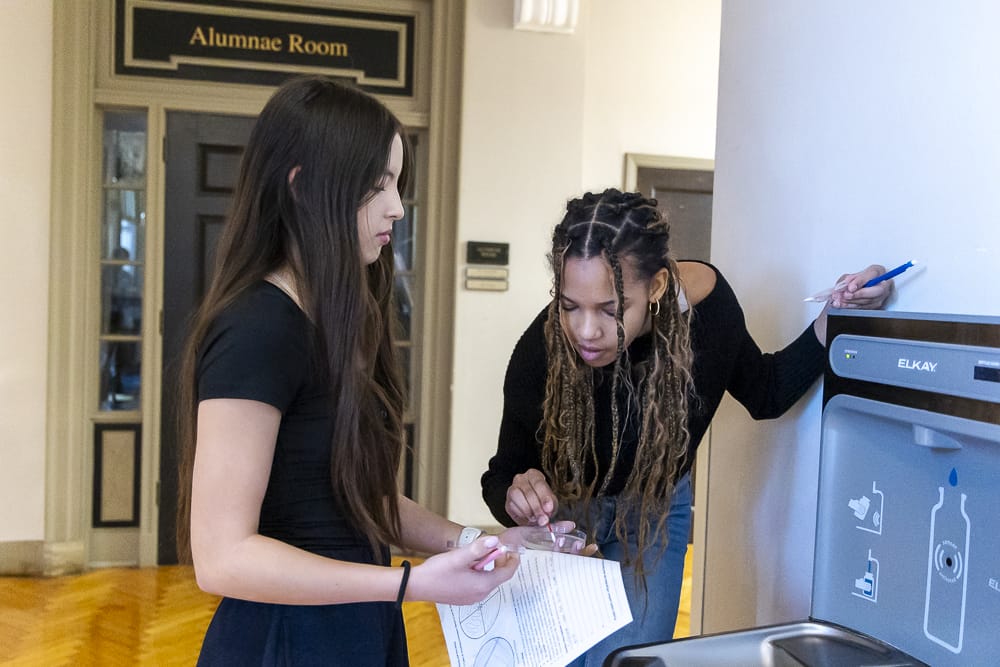
Next, students left the bacterial realm of their classroom and ventured out into the halls and rooms of the Middle School. Armed with more cotton swabs and a petri dish, they collected samples from various surfaces to investigate the presence and diversity of bacteria in their environment. They swiped the swab on a specific quadrant of the dish while trying to keep their petri dish sterile. The cell harvest location was recorded, and they had to hypothesize how the bacteria might grow.
After a few overnights of growth, the bacteria cultures were analyzed and hypotheses tested. Some bacteria didn’t form in the dishes due to sampling conditions or a non-optimum growth environment, while others produced growth from swabbed areas such as the floor and water fountain.
DNA Extraction
All living things contain DNA, and different organisms produce different DNA. Many organisms are diploids, with two copies of each chromosome. Bander chose farm-grown strawberries for the DNA lab as they are octoploids, and plant DNA tends to come from multiple sources. The students prepped their lab by smashing strawberries and preparing the solutions, which allowed the DNA to float to the top. They added a drop of the floating DNA to their slide preparation before examining the different strands through the microscope. It led to some “wow” moments of discovery and a fragrant, strawberry-scented classroom.
Kayra Metan ’29 said, “I enjoyed learning about the little things in our world that we can not see without a microscope. I was most surprised by the DNA that we extracted and how it was translucent. I was fascinated by how the DNA looked under the microscope and how it was slimy.”
Prashanti Mamileti ’30 enjoyed the variety of hands-on activities. “While learning facts was definitely fascinating, I tend to enjoy the hands-on activities as I enjoy participating! I found patience to be the most challenging part of the microscopy course. Even as I enjoyed doing things myself and getting to handle my own challenges, things such as being able to keep trying to figure out how to find bacteria became difficult. Taking this course greatly increased my respect for biologists and chemists, as they truly demonstrate optimism and patience in their work,” she said.
This engaging class bridged the gap between lab life and real-world applications, such as controlling the spread of infections and discovering patterns in DNA. What an enriching way to collaborate on scientific investigations!


